The National Museum at Bloemfontein was approached. As
this RE technology is still in the developing stage locally, medical applications
are difficult to come by. This fossil related case study was selected,
as there is very little chance that the fossil may be damaged during the
process. The electronic data as well as the final product, a prototype,
will also be used to verify this archaeological finding. This buck fossil
was found in the Kango caves near Oudtshoorn, Western Cape.
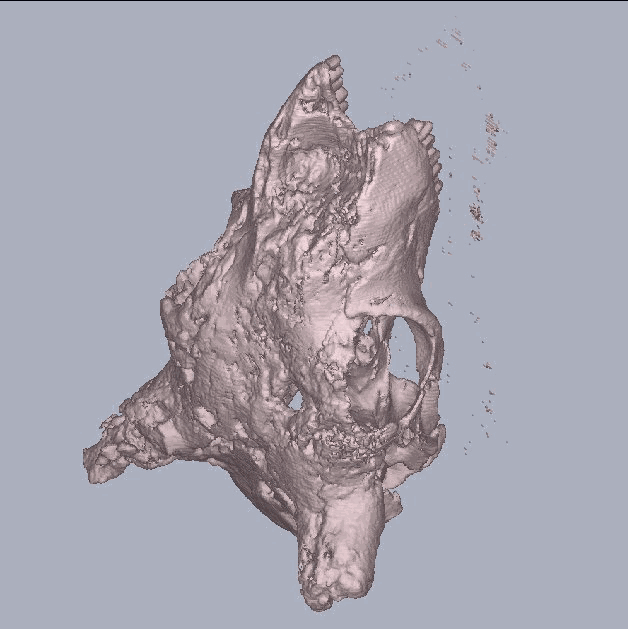
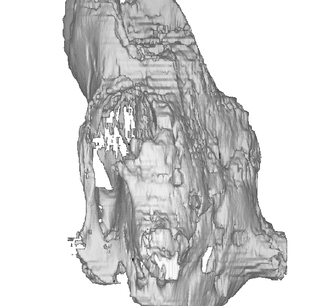
This fossil was scanned twice. The first time the fossil was scanned, the data was reconstructed at 3mm thick slice and 3mm thick intervals. This resulted in a staircase effect and poor surface finish. A great deal of data was also lost due to the coarse scanning reconstruction, low bone density and thin bone sections . Mr. Brink requested that we look into this problem as it was not acceptable to prove the archaeological finding. The second scan was reconstructed to 1mm thick slices and 1mm intervals. This images clearly show better results. There still are detail missing, most probably due to the thin bone sections and low bone density. More experimenting may need to be done to fine-tune the CAT scanner and settings to achieve the optimum conditions. An alternative method of capturing this data may be the DIGIBOTICS 4-axis laser scanner.
The data was stored in three sets. The radiographer in sets of about 45 slices stored data. This was done to allow the X-ray tube to cool down during scanning and storing stages. It also allowed a time delay for the computer to process the data. The data was stored in on the hard drive in different directories and processed separately. Surface files were generated (.srf). The various .srf files could not be joined at a later stage for 3D image processing, as they were stored indifferent directories. Moving the files into one directory did not solve the problem. The original CAT 2D images were copied to a new directory where it was converted and processed the data again, to view it as one entity.
4.3.2.1 Images
 |
 |
| Figure 4.3.2.1
3D Reconstruction of the Kango Fossil |
Figure 4.3.2 .2
3D Reconstruction of the Kango Fossil |
4.3.3 Kango Cave Buck
Data Sheet:
| Description | Options (Default) | Data | |
| 1 | CT Image Names | E6282S10I*.CT | |
| 2 | Patient/Project Name | 2S10I.pat, kbok.pat | |
| 3 | Number of First Input Image | 1/21 | |
| 4 | Number of Last Input Image | 288/306 | |
| 5 | Number of First Output Image | 000 | |
| 6 | CT or MRI | CT, MRI | CT |
| 7 | Horisontal Nr. Of Image Pixels | 0 to 65535 (265,512,1024) | 512 |
| 8 | Vertical Nr. Of Image Pixels | 0 to 65535 (265,512,1024) | 512 |
| 9 | Number of Images per File | (1) | 1 |
| 10 | File Swap Format (0,3) | 0,3 | CCGEADVA |
| 11 | Pixel Type | B,UB,S,US,L,UL,F | - |
| 12 | Header Size | *see formula below | - |
| 13 | Inter Image Header Size | 0 | - |
| 14 | Add Value | 0 to 4095 | - |
| 15 | Scale Value | 0 to 4095 | - |
| 16 | Table Position | (mm) | 0 |
| 17 | Distance Between Slices | (mm) | 1 |
| 18 | Slice thickness | (mm) | 1 |
| 19 | Pixel Size SQ. | F.O.R./Nr. Hor. Pixels (mm) | 0.47 |
| 20 | Gantry Tilt Angle | Degrees | 0 |
| 21 | Field of Reconstruction/View | (mm) | 240 |
| 22 | Number of Images | 288 | |
| 23 | File Size of CAT Image | kb | 270-280 |
| 24 | File Size of Converted Image | kb | 20-35 |
| 25 | .3dd file size | Mb | 10.56 (.srf) |
| 26 | .STL file size | Mb | - |
| 27 | RP Method | (SLA,FDM,OTHER) | - |
| 28 | .IGS file size | Mb | - |
| 29 | RP Slice file size | Mb | - |
| 30 | RP Download File size | Mb | - |
| 31 | Grow Time | Hour | - |
| 32 | Tip size | (T12, T25) | - |
| 33 | Slice Thickness | (0.01", 0.014") | - |
| 34 | Finishing Time | Hour | - |
| 35 | Processing Time | Hour | 3 |
| 36 | Data Retrieval Time | Hour | 4 |
| 37 | Total Cost | Rand | =2*7*150+1166
=3266 |
