4.14 Taung
Child Skull - Fossil
4.14.1 Background
Australopithecus africanus
TIME: Tertiary (Late Pliocene)
LOCALITY: Africa (Ethiopia, Kenya, South Africa and Tanzania)
SIZE: 4 ft 4 in/1.3 m tall
The skull of an infant specimen of A. Africanus--"southern
ape of Africa"--was unearthed in the Transvaal in 1924. It was largely
disregarded by anthropologists of the time, who thought that human origins
lay with another fossil found some years earlier in Piltdown in southern
England. This latter fossil, which appeared to possess a large brain, was
later proved a fake. A. africanus is now rightly regarded as a hominid;
one which lived from about 3 million to about 1 million years ago. Even
if A. africanus was not our direct ancestor, it was certainly very close.
The brain was small by modern standards, being about the same size as a
chimpanzee's (up to 400 cc), and the face still had heavy, apelike jaws.
The canines were reasonably large, but in other respects, the teeth were
quite human. Like A. afarensis, it was a slightly built creature, weighing
about 65lb/30kg, which walked upright. More important than its overall
appearance, though, was its lifestyle. Some have claimed that it had moved
from woodland to open savannah. Tools and cooperative hunting techniques
were already developed. To mention a few, a gang of individuals hunting
down and killing a single animal or chasing away another animals from its
kill. Others argue that the evidence is equivocal. The bone "tools" are
so ill formed that they are probably nothing more than the remains of a
hyena's meal. Although hunting was probably significant in human evolution,
it is likely that plants--including seeds, nuts, fruit, leaves, stems and
roots--formed the major part of the diet.[60]
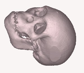
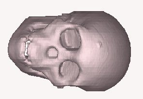
The skull of a the Taung child fossil was used in the
process to evaluate the system further. This fossil consists of three parts,
the brain area, the front facial section and part of the lower jaw. Anthropologists
used this fossil to reconstruct a complete skull of the Taung child. The
reconstructed skull and jaw were also scanned in order to capture the geometry.
Hydromed Hospital at Bloemfontein scanned these parts.
The data was retrieved and converted according to the data retrieval procedure.
4.14.2 Conclusion
The nature of the geometry is extremely complex and free
form. The complete skull featured many undercuts and hard to reach areas.
No conventional 3- or 4-axis scanning system would be used to capture this
geometry successfully. CAT scanning is the only method to reverse engineer
these parts. The x-rays penetrated the hard to reach areas and undercut
sections very easily and cost effectively. Again accuracy was not of great
importance which made this reverse engineering technique ideal to use.
4.14.2.1 Images
 |
  |
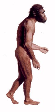
| Figure 4.14.2.1
Representation of the Australopithecus Africanus
|
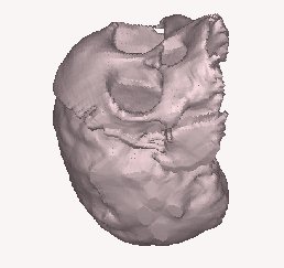
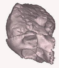
Figure 4.14.2.2
3D Images of the Taung Child fossil’s skull |
 |
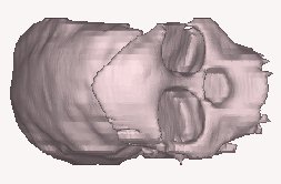 |
| Figure 4.14.2.3
The three parts of the Taung Child fossil
|
Figure 4.14.2.4
CAT scanned 3D reconstructed image
|
 |
 |
| Figure 4.14.2.5
3D image of the complete reconstructed skull |
Figure 4.14.2.6
Another representation of the reconstructed skull |
4.14.3 Taung Child Skull
Data Sheet:
| |
Description |
Options (Default) |
Data
|
| 1 |
CT Image Names |
|
Taung.00 |
| 2 |
Patient/Project Name |
|
Taung.pat |
| 3 |
Number of First Input Image |
|
34.5 |
| 4 |
Number of Last Input Image |
|
133.5 |
| 5 |
Number of First Output Image |
|
000 |
| 6 |
CT or MRI |
CT, MRI |
CT |
| 7 |
Horisontal Nr. Of Image Pixels |
0 to 65535 (265,512,1024) |
512 |
| 8 |
Vertical Nr. Of Image Pixels |
0 to 65535 (265,512,1024) |
512 |
| 9 |
Number of Images per File |
(1) |
1 |
| 10 |
File Swap Format (0,3) |
0,3 |
0 |
| 11 |
Pixel Type |
B,UB,S,US,L,UL,F |
GEADVA |
| 12 |
Header Size |
*see formula below |
- |
| 13 |
Inter Image Header Size |
0 |
- |
| 14 |
Add Value |
0 to 4095 |
- |
| 15 |
Scale Value |
0 to 4095 |
- |
| 16 |
Table Position |
(mm) |
34.5 |
| 17 |
Distance Between Slices |
(mm) |
3 |
| 18 |
Slice thickness |
(mm) |
3 |
| 19 |
Pixel Size SQ. |
F.O.R./Nr. Hor. Pixels (mm) |
0.36 |
| 20 |
Gantry Tilt Angle |
Degrees |
0 |
| 21 |
Field of Reconstruction/View |
(mm) |
185 |
| 22 |
Number of Images |
|
33 |
| 23 |
File Size of CAT Image |
kb |
270-285 |
| 24 |
File Size of Converted Image |
kb |
11-55 |
| 25 |
.3dd file size |
Mb |
1.2,1.4 |
| 26 |
.STL file size |
Mb |
9.4, 12.5 |
| 27 |
RP Method |
(SLA,FDM,OTHER) |
- |
| 28 |
.IGS file size |
Mb |
- |
| 29 |
RP Slice file size |
Mb |
- |
| 30 |
RP Download File size |
Mb |
- |
| 31 |
Grow Time |
Hour |
- |
| 32 |
Tip size |
(T12, T25) |
- |
| 33 |
Slice Thickness |
(0.01", 0.014") |
- |
| 34 |
Finishing Time |
Hour |
- |
| 35 |
Processing Time |
Hour |
- |
| 36 |
Data Retrieval Time |
Hour |
- |
| 37 |
Total Cost |
Rand |
=33*100=3300 |

Reverse Engineering Case Studies
Shortcuts back to:
Medical
Studies or
Industrial
& Biological Studies or
Fossil
Studies
or (Back to the Home Page)