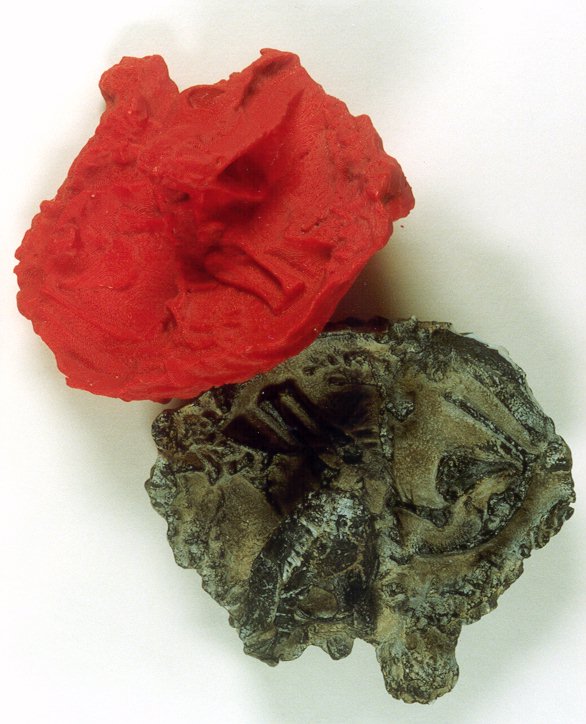
The Thrinaxodon Fossil (grey) and FDM Prototype (red)
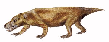
The Thrinaxodon
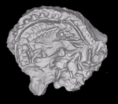
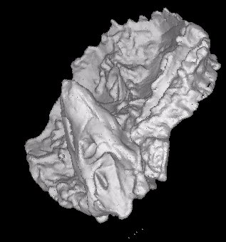
3D Reconstruction of CAT scanned fossil
3D Reconstruction of CAT scanned fossil
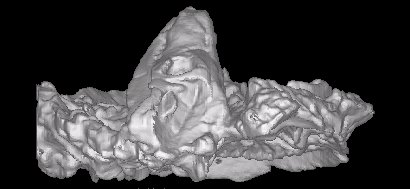
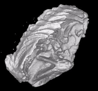
3D Reconstruction of CAT scanned fossil
3D Reconstruction of CAT scanned fossil
Thrinaxodon - A late Permian mammal-like reptile that was coming very close to being a mammal. It was a carnivorous little creature that would have grown to about the size of a small dog. Its needle-sharp teeth are classic indications of its meat diet, being present in even the smallest (youngest) of specimens. It has been suggested that Thrinaxodon was an insect-eater as well as a scavenger and a hunter of smaller animals. This interesting animal was a burrower; many fossils have been found of Thrinaxodon that died in their sleep when their tunnels obviously collapsed on them. There is disagreement between South African and American scientists about reconstruction’s of Thrinaxodon. Americans persist in putting fur on them, while South African scientists are adamant that fur had not evolved as a body covering 230-million years ago when these creatures were running around. American scientists have also speculated that Thrinaxodon were territorial animals that scent-marked their territories. Pronunciation guide: Thrinaxodon (Thry-NAKS-oh-don). Both Lystrosaurus and Thrinaxodon are creatures that inhabited the ancient Karoo area of South Africa. [15]
A plastic resin print was made from the original fossil. A great deal of rock was still inside the fossil. This rock is in general not removed as it is too difficult and not easily accessible. The resin fossil was CAT scanned at Krugersdorp Private Hospital. The data was retrieved and converted according to the procedure earlier mentioned.
This particular case study was a real treat. The data was converted with great ease in a very short period. The next stage, converting into 3D imaging was also rapid and effortless. A 3D model was created and a STL model was exported with the aid of the Materialise CTM software.
The STL file was imported into the QuickSlice software and processed.
The processing took place on a Silicon Graphics Indy computer, called INDY1.
The data processing failed miserably, after two chances. The computer just
could not manage the large data files. The Quick Slice software was installed
on the Silicon Graphics O2 computer, called LANCRE. Various grow parameters
were selected and the processing was started again. This computer processed
successfully, generating the horizontal slice, SSL file as well as the
machine language, SML file. The SML file was then transferred, via our
LAN with the aid of FTP, to INDY1 as it was connected directly to the FDM
machines. The prototype was removed from the working envelope and the support
structure was removed and cleaned.
This is the ideal method for capturing complex, low tolerance and detailed
geometry. A FDM prototype was grown to verify the process. The RE method
was used to get the data in an electronic format so that it can be used
in other processes.
|
|
| Figure 4.2.2.1
The Thrinaxodon Fossil (grey) and FDM Prototype (red) |
Figure 4.2.2.2
The Thrinaxodon |
|
|
| Figure 4.2.2.3
3D Reconstruction of CAT scanned fossil |
Figure 4.2.2.3
3D Reconstruction of CAT scanned fossil |
|
|
|
| Figure 4.2.2.3
3D Reconstruction of CAT scanned fossil |
Figure 4.2.2.3
3D Reconstruction of CAT scanned fossil |
| Description | Options (Default) | Data
|
|
| 1 | CT Image Names | bpi2.001 | |
| 2 | Patient/Project Name | bpi2.pat | |
| 3 | Number of First Input Image | 1 | |
| 4 | Number of Last Input Image | 99 | |
| 5 | Number of First Output Image | 000 | |
| 6 | CT or MRI | CT, MRI | CT |
| 7 | Horisontal Nr. Of Image Pixels | 0 to 65535 (265,512,1024) | 512 |
| 8 | Vertical Nr. Of Image Pixels | 0 to 65535 (265,512,1024) | 512 |
| 9 | Number of Images per File | (1) | 1 |
| 10 | File Swap Format (0,3) | 0,3 | CCELSC |
| 11 | Pixel Type | B,UB,S,US,L,UL,F | - |
| 12 | Header Size | *see formula below | - |
| 13 | Inter Image Header Size | 0 | - |
| 14 | Add Value | 0 to 4095 | - |
| 15 | Scale Value | 0 to 4095 | - |
| 16 | Table Position | (mm) | 0 |
| 17 | Distance Between Slices | (mm) | 1 |
| 18 | Slice thickness | (mm) | 1.2 |
| 19 | Pixel Size SQ. | F.O.R./Nr. Hor. Pixels (mm) | 0.47 |
| 20 | Gantry Tilt Angle | Degrees | 0 |
| 21 | Field of Reconstruction/View | (mm) | 240 |
| 22 | Number of Images | 99 | |
| 23 | File Size of CAT Image | kb | 75 |
| 24 | File Size of Converted Image | kb | 60 |
| 25 | .3dd file size | Mb | 25 |
| 26 | .STL file size | Mb | 26.75 |
| 27 | RP Method | (SLA,FDM,OTHER) | FDM |
| 28 | .IGS file size | Mb | - |
| 29 | RP Slice file size | Mb | 9 |
| 30 | RP Download File size | Mb | 10.8 |
| 31 | Grow Time | Hour | 41.4 |
| 32 | Tip size | (T12, T25) |
|
| 33 | Slice Thickness | (0.01", 0.014") | 0.01" |
| 34 | Finishing Time | Hour | 4 |
| 35 | Processing Time | Hour | 2 |
| 36 | Data Retrieval Time | Hour | 4 |
| 37 | Total Cost | Rand | =41.4*100+6*150+4*55+500
=5760 |
