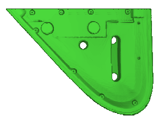
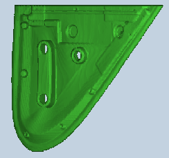
3D reconstructed image of the motorcar door window rubber.
3D reconstructed motorcar door window rubber.

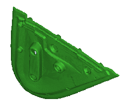
3D reconstructed motorcar door window rubber.
3D reconstructed motorcar door window rubber.
Once again the geometry was required that was used to
produce injection mould tooling. The rubber seal was taken to Krugersdorp
Hospital to capture the geometry. One millimeter thick slices were required
to obtain sufficient detail of the seal’s geometry. Three data sets of
sixty slices each were made due to the maximum amount of slices that can
be scanned at one time. The limitations are related to the computer processing
capabilities, hard drive space and X-ray tube cools down periods. The seal
was aligned in two axis with the visual aids available on the CAT scanner.
A flat section of foam was used to suspend and separate the seal from the
scanner’s table. This would help in later during the data conversion and
processing stages. CAT scanning took place at 130kV.
The threshold, type of material and material cross sectional wall thickness here plays a cardinal role. One can easily adjust the threshold to vary the wall or cross section. Any imperfection will also be captured with this method. A fair amount of editing can be applied to solve this problem. If the material cross section is to thin or if the material density is too low, a poor CAT image will be generated. To improve the poor data that was captured, the threshold can be increased. The threshold can be increased to such an extent that the 2mm-wall thickness can be thickened to 3.5mm. It is therefor important that some form of reference or verification is implemented every time. It is also required by the ISO 9000/1 standards, that results and processes can by traced and verified. It was again clear that the correct tools are still not in place at the TCT, CSIR, to perform the tasks. STL data manipulation tools are requited that can perform boolean operations, section 3D STL geometry, fix and verify STL data, tools that can generate machine tool paths from STL data. STL format is one of the easiest data format obtainable from RE, and it is therefore important to have the correct software programs that can manipulate STL format files.
4.9.2.1. Images
| Description | Options (Default) | Data
|
|
| 1 | CT Image Names | mirror.00 | |
| 2 | Patient/Project Name | spiel.pat | |
| 3 | Number of First Input Image | mirror1.000 @ 34.5 | |
| 4 | Number of Last Input Image | mirror1.147 @ 179.8 | |
| 5 | Number of First Output Image | 000 | |
| 6 | CT or MRI | CT, MRI | CT |
| 7 | Horisontal Nr. Of Image Pixels | 0 to 65535 (265,512,1024) | 512 |
| 8 | Vertical Nr. Of Image Pixels | 0 to 65535 (265,512,1024) | 512 |
| 9 | Number of Images per File | (1) | 1 |
| 10 | File Swap Format (0,3) | 0,3 | CCELSC |
| 11 | Pixel Type | B,UB,S,US,L,UL,F | - |
| 12 | Header Size | *see formula below | - |
| 13 | Inter Image Header Size | 0 | - |
| 14 | Add Value | 0 to 4095 | - |
| 15 | Scale Value | 0 to 4095 | - |
| 16 | Table Position | (mm) | 34.5 |
| 17 | Distance Between Slices | (mm) | 1 |
| 18 | Slice thickness | (mm) | 1.2 |
| 19 | Pixel Size SQ. | F.O.R./Nr. Hor. Pixels (mm) | 0.684 |
| 20 | Gantry Tilt Angle | Degrees | 0 |
| 21 | Field of Reconstruction/View | (mm) | 350 |
| 22 | Number of Images | 147 | |
| 23 | File Size of CAT Image | kb | 27 |
| 24 | File Size of Converted Image | kb | 19-22 |
| 25 | .3dd file size | Mb | 0.234 |
| 26 | .STL file size | Mb | 8.9 |
| 27 | RP Method | (SLA,FDM,OTHER) | - |
| 28 | .IGS file size | Mb | - |
| 29 | RP Slice file size | Mb | - |
| 30 | RP Download File size | Mb | - |
| 31 | Grow Time | Hour | - |
| 32 | Tip size | (T12, T25) | - |
| 33 | Slice Thickness | (0.01", 0.014") | - |
| 34 | Finishing Time | Hour | - |
| 35 | Processing Time | Hour | 1 |
| 36 | Data Retrieval Time | Hour | 3 |
| 37 | Total Cost | Rand | 600+800=1200 |
