-
4.12 Maxilla Cranio Facial – Medical
This case study again shows the necessity of rapid prototyping
in medicine.
4.12.1 Background
The patient, a three-year-old boy, was scanned at the
Pretoria East Hospital clinic with the aid of their Toshiba Spiral X-Vision/GX
CAT scanner. The CAT scanner at Pretoria East Hospital based in Pretoria.
The 2D CAT slices were again stored on ¼" data cartridge magnetic
tape. 2D CAT images of various positions were stored on film for record.
The patient required maxilla cranio facial surgery.
Maxilla-Facial specialists from the University of Pretoria
requested a prototype of the patient’s skull. The skull was required for
pre-surgical planning.
Permission was granted so that this patient’s data may
be retrieved from the CAT scanner. Please refer to section (3.7) for the
data retrieval procedures. Pretoria East Hospital prepared a 3D visualization
of the scanned area. The spiral data was prepared in one set. The radiology
team prepared one set of 3mm thick slices and 3mm intervals or spacing.
The finest possible slice thickness and slice spacing were again requested
to obtain the best results. In this case it would be better to sample more
data than required. The CAT scanner parameters were restricted to scanning
distance and slice thickness. The surgeon was particularly interested in
the frontal or facial area of the skull. For a successful facial reconstruction,
the facial geometry had to be known.
The data was then processed and a meeting was arranged
with the surgeons, to verify the results on the computer screen. They were
please with the results and requested that we continue with the next phase
of the project.
The next stage was to create a prototype of the skull.
The 3dd file was used with the Materialise, CTM software to generate a
STL format file. The STL file was then used with the Stratasys QuickSlice
software. Horizontal slices were created. The software performed support
generation, automatically. The SML file was created. The FDM machines require
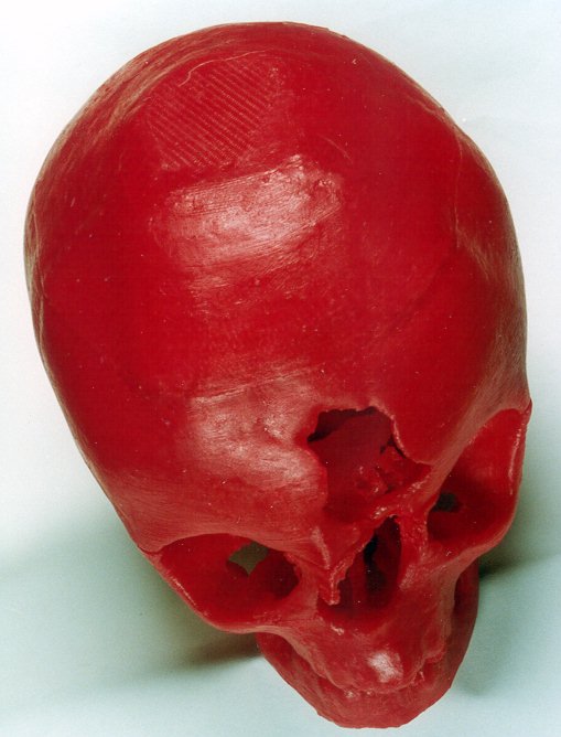
this data to build a prototype. The prototype was grown with the red ICW06
wax material and finished by removing the support structure generated by
the FDM process. The skull was grown in two section to simplify the prototyping
process. This also made the removal of the support structure easier.
The prototype of the skull was supplied to the maxilla-facial
surgeons. This prototype could only be used for visual inspection. Wax
prototypes are very fragile and can break easily when handled. It also
does not machine easily. A bronze casting was made from this wax skull
with the aid of the investment casting process.
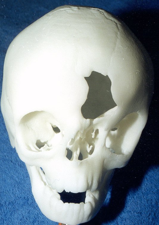
A second skull prototype was produced by a group in Holland.
The South African Selective Laser Sintering (SLS) rapid prototyping system
was not operational at that stage. The skull was produced in the glass
filled nylon material.
4.12.2 Conclusion
The SLS system proved to be the best system to use with
this technology. It is of great use to produce prototypes without the generation
of a support structure. The support structure always need to be removed
and very often causes problems to the extent of damaging the prototype.
The surgeons worked with ease with the glass filled nylon
material prototype. The skull prototype was marked and cut in sections
as the surgeons planned the operation. A brace was also fitted to reposition
the eye socket section.
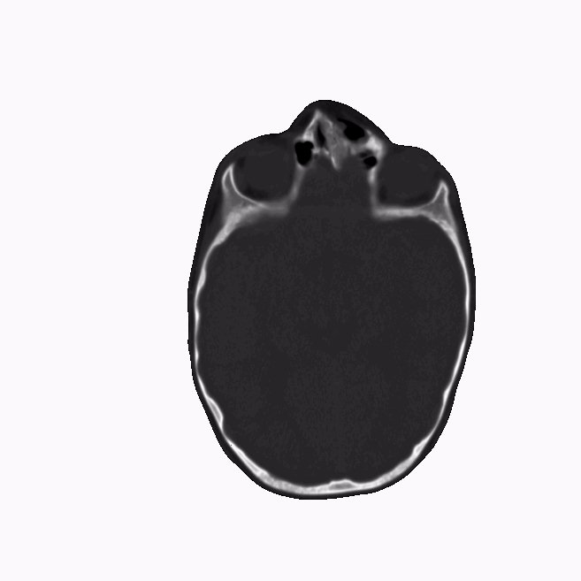
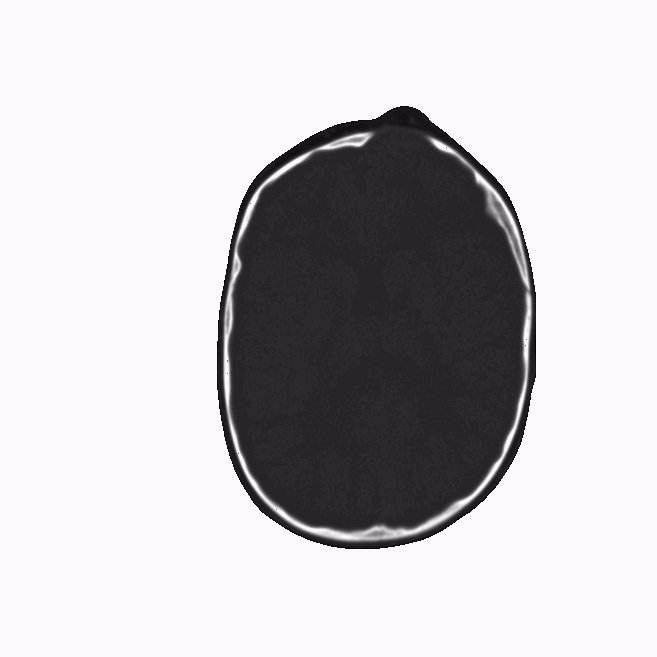
2D CAT images were checked during scanning. . CAT scanned
film was also provided with 2D slice information as well as 3D rendering
to be used for verification. The window settings were optimized on the
CT computer. Measurements were checked. Window and threshold settings were
set to the ideal conditions.
Both the maxilla cranio- facial- and neuro-surgeons frequently
referred to the skull prototype during the eight-hour operation.
The Bronze casting proved to be useful for preserving
the wax prototype but unfortunately warped slightly. Other casting imperfections
such as pit holes were evident.
This skull data was supplied to the CSIR Virtual Reality
Centre. Initially they battled to retrieve the data due to a lack of computer
power. This skull is now used to demonstrate the virtual reality surgery
concept to be used by medical students.
4.12.2.1 Images
 |
 |
| Figure 4.12.2.1
2D CAT scan slice at the 1100 position |
Figure 4.12.2.2
2D CAT scan slice at the 1080 position
|
 |
 |
| Figure 4.12.2.3
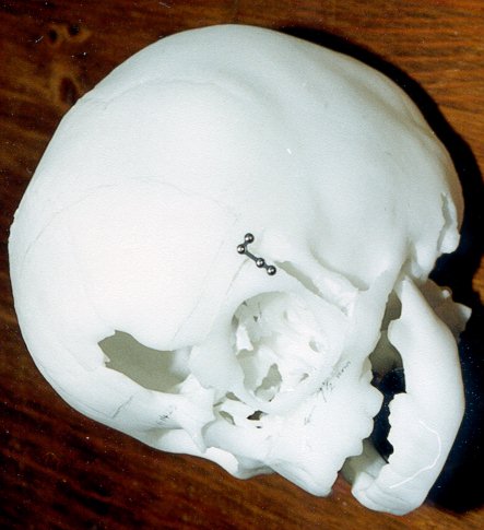
SLS skull prototype in GF nylon
|
Figure 4.12.2.4
Bronze casting of the skull
|
 |
 |
| Figure 4.12.2.5
FDM skull prototype in ICW06 wax.
|
Figure 4.12.2.6
Surgical Reconstructed SLS prototype.
|
 |
 |
| Figure 4.12.2.7
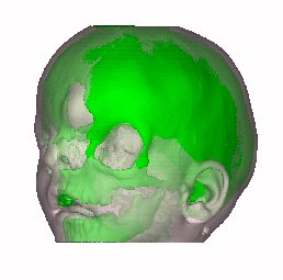
3D Image of skull, combines soft and hard tissue. |
Figure 4.12.2.8
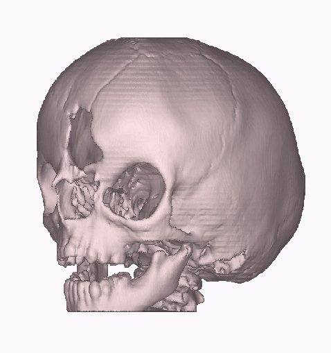
3D Image of skull, bone structure only.
|
4.12.3 Maxilla Cranio Facial Data Sheet:
| |
Description |
Options (Default) |
Data
|
| 1 |
CT Image Names |
|
seun.00 |
| 2 |
Patient/Project Name |
|
seun.pat |
| 3 |
Number of First Input Image |
|
1.6912.2 |
| 4 |
Number of Last Input Image |
|
1.6912.319 |
| 5 |
Number of First Output Image |
|
seun.00 |
| 6 |
CT or MRI |
CT, MRI |
CT |
| 7 |
Horisontal Nr. Of Image Pixels |
0 to 65535 (265,512,1024) |
512 |
| 8 |
Vertical Nr. Of Image Pixels |
0 to 65535 (265,512,1024) |
512 |
| 9 |
Number of Images per File |
(1) |
1 |
| 10 |
File Swap Format (0,3) |
0,3 |
Toshiba |
| 11 |
Pixel Type |
B,UB,S,US,L,UL,F |
- |
| 12 |
Header Size |
*see formula below |
- |
| 13 |
Inter Image Header Size |
0 |
- |
| 14 |
Add Value |
0 to 4095 |
- |
| 15 |
Scale Value |
0 to 4095 |
- |
| 16 |
Table Position |
(mm) |
-1021.5 |
| 17 |
Distance Between Slices |
(mm) |
3 recon to 1 |
| 18 |
Slice thickness |
(mm) |
3 recon to 1 |
| 19 |
Pixel Size SQ. |
F.O.R./Nr. Hor. Pixels (mm) |
0.39 |
| 20 |
Gantry Tilt Angle |
Degrees |
0 |
| 21 |
Field of Reconstruction/View |
(mm) |
200 |
| 22 |
Number of Images |
|
154 |
| 23 |
File Size of CAT Image |
kb |
521 |
| 24 |
File Size of Converted Image |
kb |
76-131 |
| 25 |
.3dd file size |
Mb |
6*.srf |
| 26 |
.STL file size |
Mb |
96, 17.9 with filter |
| 27 |
RP Method |
(SLA,FDM,OTHER) |
FDM, SLS |
| 28 |
.IGS file size |
Mb |
- |
| 29 |
RP Slice file size |
Mb |
28.9+19.4 |
| 30 |
RP Download File size |
Mb |
12.1+13.9 |
| 31 |
Grow Time |
Hour |
132.9 |
| 32 |
Tip size |
(T12, T25) |
T12 |
| 33 |
Slice Thickness |
(0.01", 0.014") |
0.01" |
| 34 |
Finishing Time |
Hour |
8 |
| 35 |
Processing Time |
Hour |
16 |
| 36 |
Data Retrieval Time |
Hour |
3 |
| 37 |
Total Cost |
Rand |
=27*250+7500=14250 |

Reverse Engineering Case Studies
Shortcuts back to:
Medical
Studies or
Industrial
& Biological Studies or
Fossil
Studies
or (Back to the Home Page)