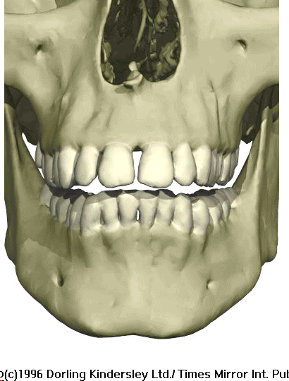
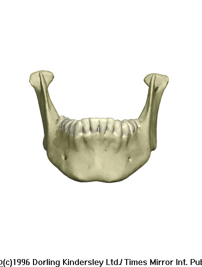
The Human Maxilla and Mandible.
The Human Mandible.

The Human Mandible.
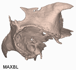
The patient’s reconstructed Maxilla.
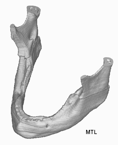
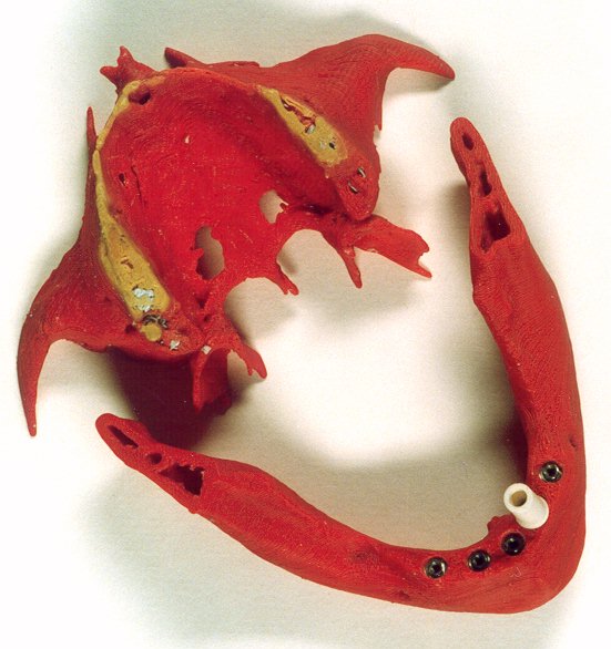
The patient’s reconstructed Mandible.
FDM Prototypes of the patient’s Mandible and Maxilla with dental implants fitted.
The maxillary bone, or maxilla, of the skull holds the upper teeth and encloses the opening for the nasal passage. The upper part of the bone meets the parietal and nasal bone between the eyes. The maxilla also meets the zygomatic bone (cheekbone) below the eye. The lower part of the maxilla has sockets for the eight teeth of one side of the upper jaw. Behind this, the maxilla also forms the front part of the roof of the mouth, or hard palate, which creates a firm resistance when the tongue is manipulating food in the mouth. This palate also separates the mouth from the air passage that leads from the nose to just above the windpipe at the back of the throat. [37], [36]
The mandible (jawbone) is connected to the upper part of the skull, or cranium, at a joint in the temporal bone. It contains the sockets for the 16 lower teeth. Like some of the bones of the skull, it first appears in the developing baby, or fetus, as a pair of bones, but these later fuse together to make the mandible stronger.
The mandible has to be strong, since strong muscles are connected to it. The temporal muscle is attached to part of the front of the jaw joint, known as the coronoid process. The masseter muscle is attached to the angle of the jaw. You use both of these muscles when you close the mouth or chew. The mouth is opened by using the digastric muscle. It is connected to the back of the mandible under the chin, and the lateral pterygoid (pronounced "terrygoid") muscle, which runs from the inner side of the jaw to the underside of the skull.
Adults have eight teeth in each quarter of the mouth - two incisor teeth, one canine tooth, two premolar teeth and three molar teeth, making a total of 32 in the whole of the mouth. This adult set of teeth is unlike our first set of baby teeth, called "milk teeth." Milk teeth do not include any molar teeth, so children only have five teeth in each quarter of the mouth, and therefore have only 20 teeth altogether.
Both adult and milk teeth are sunk into sockets in the bones that hold them - the upper teeth are held in the maxilla (a bone of the skull), and the lower teeth are held in the mandible (jawbone). The roots of the teeth are up to twice as long as the part of the tooth that can be seen above the gum.
The first incisor tooth lies at the front of the mouth. Like all the teeth, it is made mostly of a substance called dentine, which is a hard tissue that is much tougher than normal bone. The first incisor has a sharp edge that is parallel with the front of the mouth, and which is used to bite through the tough parts of food. There are two incisor teeth in each quarter of the mouth, and the one closest to the middle of the jaw is larger than the outer one. Each incisor tooth has a single root.
The second incisor tooth lies near the front of the mouth. It has a sharp edge that is parallel with the front of the mouth. This tooth is used to bite through the tough parts of food. There are two incisor teeth in each quarter of the mouth, and the one closest to the middle of the jaw is larger than the outer one.
The canine teeth lie near the front of the mouth, between the incisor teeth and the premolar teeth. There is only one canine tooth in each quarter of the mouth. The end of each canine tooth is pointed, and it is used with the incisor teeth to bite through the tough parts of food. The first premolar tooth lies toward the side of the mouth, between the canine tooth and the second premolar tooth. Its surface has two rounded projections, called "cusps," and the premolar teeth help the molar teeth to chew food. The first upper premolar usually has two roots, but the lower one has only one root.
Each canine tooth has a single root.
The first molar tooth lies in the rear part of the mouth, between the second premolar tooth and the second molar tooth. It has either two or three roots. Its surface has four projections, called "cusps," that crush and chew food. Like the other two molar teeth, it lies underneath the strong jaw muscles, so it is very effective in chewing up food. The second molar tooth lies in the rear part of the mouth, between the first molar tooth and the third molar tooth. It has either two or three roots. Its surface has four projections, called "cusps," that crush and chew food. Like the other two molar teeth, it lies underneath the strong jaw muscles, so it is very effective in chewing up food.
The third molar tooth lies furthest back in the mouth
- if it appears at all during your lifetime. It develops in the skull when
the child is nine years old, but it does not appear through the gum until
the age of 17 to 21 years. It is sometimes called the "wisdom" tooth, because
it is thought that greater age brings greater wisdom. In some cases, the
third molar teeth do not ever push through the gum.
A 41-year-old female patient required a dental implant. The prostodontist was contacted and it was agreed that it will be of great value to capture the geometry of the maxilla and mandible to determine if there is sufficient bone structure required for an operation of this nature. The patient was scanned at Morningside clinic with the aid of their Toshiba spiral X-Vision/GX CAT scanner. From the spiral data, sagittal and coronal multi-planar reconstruction images were made. The CAT scanned film was used to verify the converted data. This was useful. However, the film did not represent the axial CAT scan images that was used to reconstruct the 3D data. Reference [40] p 44, recommend a 1.5mm slice thickness with a 1mm table feed. The X-ray energy should not exceed 100mA per section. The patients' head needs to be fixed securely.
Permission was granted so that this patient’s data may
be retrieved from the CAT scanner.
4.5.2 Conclusion
Delivery turn around time and cost are the most important factors to the prostodontist. He suggested that the surgeon would be very keen to obtain routine prototypes if the cost was in the region of several hundred Rand. Once a larger market is established, the cost will reduce.
Faster turns around times are also critical. This can only be achieved to have a tape streamer fitted to the CAT scanner so that data storage can take place without interrupting the workflow. This will also require a dedicated RP system. Several measurements were taken at slice 27 and the dimensions varied from 0.46mm in the z-axis to 1.48 mm in the xy-axis. The pixel size was 0.26 mm and therefor a tolerance of ± 0.26mm should be acceptable. The part in the 3D Mimics software was in both cases larger. It was very difficult to determine the accuracy as the printed 2D CAT scan images, multi-planar reconstruction, did not correlate with the axial CT images that was used to reconstruct the 3D model. The deviation may be due to the thin bone section in the maxilla. The threshold may be set too low to obtain a full maxilla 3D image, but will result in an oversize mandible.
The 2D images were processed. Two different regions were
selected, one for the maxilla and one for the mandible. Different thresholds
should also have been used for the two regions or masks to obtain optimal
accuracy.
4.5.2.1. Images
4.5.3 Maxilla and
Mandible Data Sheet:
| Description | Options (Default) | Data
Maxilla |
Data Mandible | |
| 1 | CT Image Names | cruz | cruz | |
| 2 | Patient/Project Name | cruz.pat | cruz.pat | |
| 3 | Number of First Input Image | 1:643:9/-19.5 | 1:643:9/-19.5 | |
| 4 | Number of Last Input Image | 1:643:155/53.5 | 1:643:155/53.5 | |
| 5 | Number of First Output Image | 000 | 000 | |
| 6 | CT or MRI | CT, MRI | CT | CT |
| 7 | Horisontal Nr. Of Image Pixels | 0 to 65535 (265,512,1024) | 512 | 512 |
| 8 | Vertical Nr. Of Image Pixels | 0 to 65535 (265,512,1024) | 512 | 512 |
| 9 | Number of Images per File | (1) | 1 | 1 |
| 10 | File Swap Format (0,3) | 0,3 | 0 | 0 |
| 11 | Pixel Type | B,UB,S,US,L,UL,F | S | S |
| 12 | Header Size | *see formula below | 8704 | 8704 |
| 13 | Inter Image Header Size | 0 | 0 | 0 |
| 14 | Add Value | 0 to 4095 | 2048 | 2048 |
| 15 | Scale Value | 0 to 4095 | 1.8 | 1.8 |
| 16 | Table Position | (mm) | 0 | 0 |
| 17 | Distance Between Slices | (mm) | 0.5 | 0.5 |
| 18 | Slice thickness | (mm) | 3 | 3 |
| 19 | Pixel Size SQ. | F.O.R./Nr. Hor. Pixels (mm) | 0.2598 | 0.2598 |
| 20 | Gantry Tilt Angle | Degrees | 0 | 0 |
| 21 | Field of Reconstruction/View | (mm) | 133 | 133 |
| 22 | Number of Images | 146 | 146 | |
| 23 | File Size of CAT Image | kb | 252 | 252 |
| 24 | File Size of Converted Image | kb | 180 | 180 |
| 25 | .3dd file size | Mb | 15 | 13 |
| 26 | .STL file size | Mb | 26.37 | 32 |
| 27 | RP Method | (SLA,FDM,OTHER) | FDM | FDM |
| 28 | .IGS file size | Mb | - | - |
| 29 | RP Slice file size | Mb | 6.79 | 3.7 |
| 30 | RP Download File size | Mb | 5.9 | 7.6 |
| 31 | Grow Time | Hour | 18.25 | 23.75 |
| 32 | Tip size | (T12, T25) | T12 | T12 |
| 33 | Slice Thickness | (0.01", 0.014") | 0.01" | 0.01" |
| 34 | Finishing Time | Hour | 8 | 6 |
| 35 | Processing Time | Hour | 6 | 6 |
| 36 | Data Retrieval Time | Hour | 2 | 2 |
| 37 | Total Cost | Rand | =1500+330+
1825 = 3655 |
=8*150+6*55+
2375 = 3905 |
